Dove Mountain Veterinary
Ultrasound
Today, ultrasound is used in the same way to detect abnormalities in the soft tissues.

When an animal has an injury or illness, new technologies can be critical for finding out what is wrong and what to do about it. Veterinarians have used x-rays to view the hard structures inside the animal’s body to find problems. Today, ultrasound is used in the same way to detect abnormalities in the soft tissues. At Dove Mountain Veterinary in Marana, AZ, we use ultrasounds technology to help detect problems and ensure accurate treatment for our patients.
FAQs
Understanding Ultrasound Technology
Ultrasounds uses sound waves that bounce against internal structures to produce an echo. This echo is then translated into a digital image on a computer screen. By studying these images, a veterinarian can detect changes in the size and shape of internal organs that might indicate disease or injury. The images, along with other types of testing, help to pinpoint the problem, so the most effective treatment can be provided.
Conditions Ultrasound Can Detect
Because ultrasound allows veterinarians a way to look inside the body without having to do invasive surgery, it has become an important tool for diagnosis. Vets use ultrasound to diagnose reproductive problems during pregnancy and birth, just as it is used in human medicine. But the technology can also be used to view the joints, the kidneys, the urinary bladder, the liver, the heart and the blood vessels. It can also help to diagnose obstructions in the intestines. Ultrasound is also used to help the vet guide needles for biopsies of internal tissues.
What Happens During An Ultrasound?
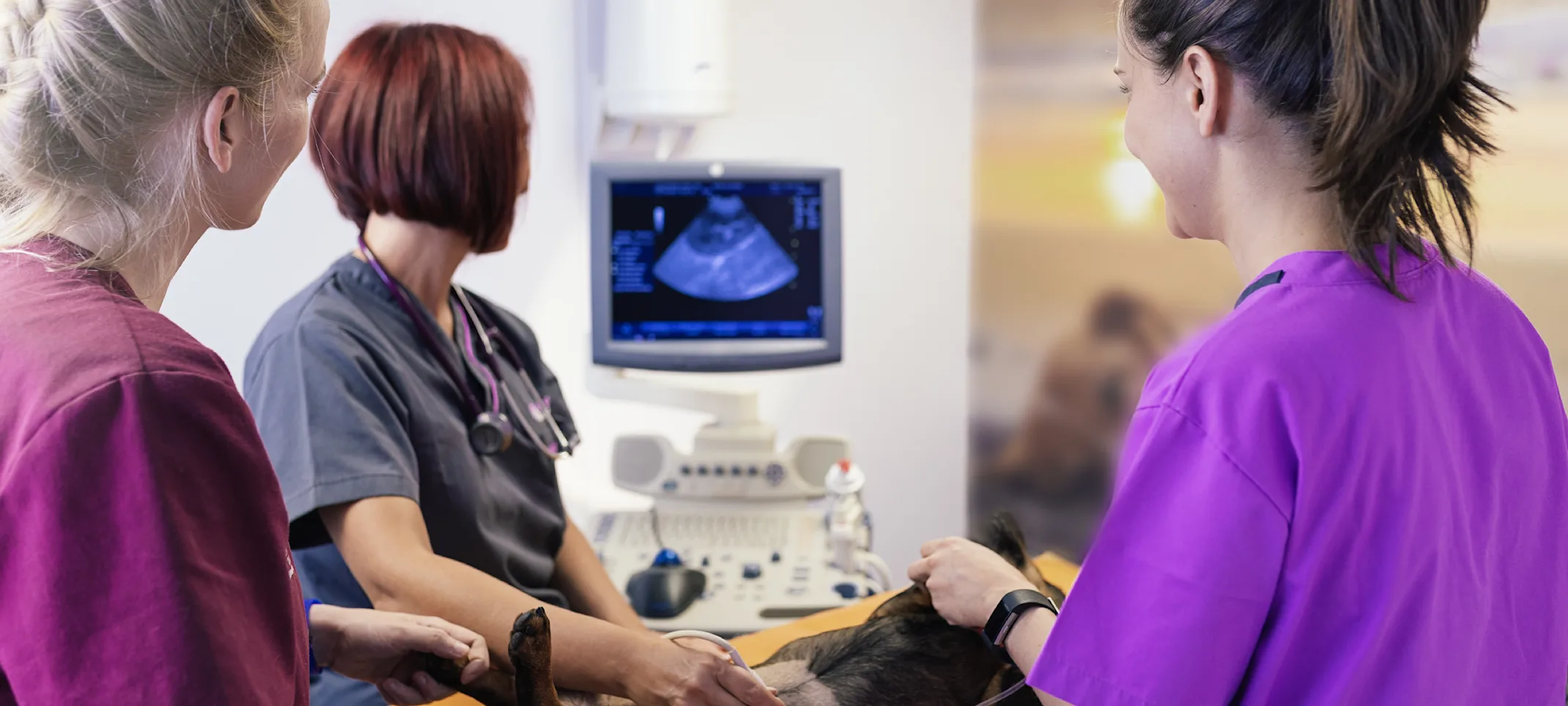
During an ultrasound test, the animal lies down on an examining table quietly. Generally, it is not necessary to administer an anesthetic. However, very active animals may be given a sedative to allow the vet to get a clear picture of internal organs. The area of the body being tested may have to be shaved. The vet uses a hand-held device that is passed over the surface of the skin to produce the computerized image. Then, the vet can analyze this image to make a diagnosis and determine the right treatment.
